
Amniotic fluid embolism-related coagulopathy should be managed with hemostatic resuscitation with the use of a 1:1:1 ratio of packed red cells, fresh frozen plasma, and platelets (with cryoprecipitate as needed to maintain a serum fibrinogen of >150-200 mg/dL). Blood pressure support with vasopressors is preferred over fluid infusion in the setting of severe right ventricular compromise. If such failure is identified, treatment that is tailored at improving right ventricular performance should be initiated with the use of inotropic agents and pulmonary vasodilators. The first English language description appeared in 1941 and consisted of eight parturients dying suddenly in which, once again, fetal material was. Where available, we recommend performing transthoracic or transesophageal echocardiography as soon as possible because this is an easy and reliable method of identifying a failing right ventricle. The embolus may be a blood clot (thrombus), a fat globule (fat embolism), a bubble of air or other gas (gas embolism), amniotic fluid (amniotic fluid embolism), or foreign material. Amniotic fluid embolism was first recognized in 1926, in a Brazilian journal case report, on the basis of large amounts of fetal material in the maternal pulmonary vasculature at autopsy.

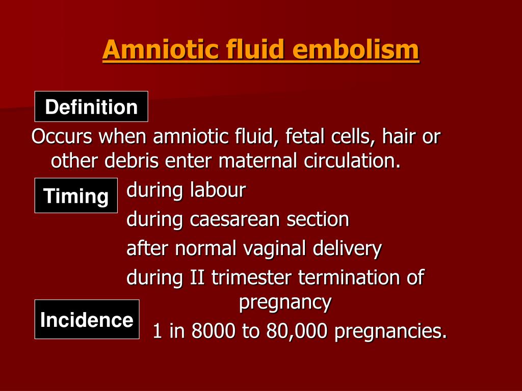
We describe key features of initial treatment of patients with amniotic fluid embolism. Because amniotic fluid embolism usually is seen with cardiac arrest, the initial immediate response should be to provide high-quality cardiopulmonary resuscitation. Amniotic fluid embolism is an uncommon, but potentially lethal, complication of pregnancy.


 0 kommentar(er)
